Thursday 24 March 2016

Detection of Helicobacter pylori antigen by enzyme immunoassay (EIA)

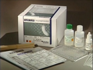
Helicobacter pylori antigen test was performed using Premier Platinum HpSA PLUS enzyme immunoassay (EIA). The kit used was purchased from Meridian Bioscience Inc. The Premier Platinum HpSA PLUS enzyme immunoassay (EIA) kit is an in vitro qualitative procedure for the detection of Helicobacter pylori antigens in human stool. Test results are intended to aid in the diagnosis of H. pylori infection and to monitor response during and post-therapy in patients. Accepted medical practice recommends that testing by any current method, to confirm eradication, be done at least four weeks following completion of therapy.
Helicobacter pylori which is previously known as Campylobacter pylori, is a gram negative, microaerophilic bacterium usually found in the stomach. It was identified in 1982 by Australian scientists Barry Marshall and Robin Warren, who found that it was present in a person with chronic gastritis and gastric ulcers, conditions not previously believed to have a microbial cause. It is also linked to the development of duodenal ulcers and stomach cancer. However, over 80% of individuals infected with the bacterium are asymptomatic, and it may play an important role in the natural stomach ecology. According to research, more than 50% of the world's population harbour H. pylori in their upper gastrointestinal tract. Infection is more prevalent in developing countries, and incidence is decreasing in Western countries. H. pylori's helical shape and its flagella is used to penetrate the mucoid lining of the stomach to reach the epithelial cells underneath, where the pH is more neutral. They also neutralise the acid in its environment by producing large amounts of urease, which breaks down the urea present in the stomach to carbon dioxide and ammonia. The ammonia, which is basic, then neutralizes stomach acid. This ammonia produced to regulate pH is toxic to epithelial cells, as well as other biochemicals produced by H. pylori such as proteases, vacuolating cytotoxin A (VacA) and certain phospholipases. Cytotoxin associated gene CagA can also cause inflammation and is potentially a carcinogen.
Symptoms
Most people infected with H. pylori are asymptomatic but in acute infection may appear as an acute gastritis with abdominal pain or nausea. If the infection develops into chronic gastritis, the symptoms may include nausea, belching, stomach pains, bloating, black stool and sometimes vomiting
Individuals infected with H. pylori have a 10 - 20% lifetime risk of developing peptic ulcers and a 1 - 2% risk of acquiring stomach cancer. Inflammation of the pyloric antrum is more likely to lead to duodenal ulcers, while inflammation of the corpus (body of the stomach) is more likely to lead to gastric ulcers and gastric carcinoma. However, H. pylori possibly plays a role only in the first stage that leads to common chronic inflammation, but not in further stages leading to carcinogenesis. A meta-analysis conducted in 2009 concluded the eradication of H. pylori reduces gastric cancer risk in previously infected individuals, suggesting the continued presence of H. pylori constitutes a relative risk factor of 65% for gastric cancers and in terms of absolute risk, the increase was from 1.1 - 1.7%. H. pylori have been associated with colorectal polyps and colorectal cancer and may also be associated with eye disease
SPECIMEN COLLECTION AND PREPARATION
The faeces sample should be received in an airtight transport container and stored at 2-8oC until tested. The specimen should be tested as soon as possible, but may be held up to 72 hours at 2-8oC prior to testing. If testing cannot be performed within this time frame, specimens should be frozen immediately upon receipt and stored frozen (-20oC to –80oC) until tested. Specimens may be frozen and thawed twice.
Stool in transport media, swabs, or preservatives are inappropriate for testing.
SPECIMEN PREPARATION
1. Using a pipetting device, add 500μL of Sample Diluent to a clean test tube.
2. Mix stool as thoroughly as possible prior to pipetting.
a. Liquid or semi-solid stools - Using the supplied transfer pipette, add 100μL (second mark from the tip of the pipette) of stool into Sample Diluent. Using same pipette, gently withdraw and expel the stool suspension several times, then vortex 15 seconds. Save the transfer pipette in the sample for later use.
b. Formed/Solid stools - Using a wooden applicator stick, transfer a small portion (5-6 mm diameter) of thoroughly mixed stool into Sample Diluent. Emulsify stool using the wooden applicator stick, then vortex 15 seconds.
3. Stool specimens may be centrifuged after dilution. Centrifuge at approximately
2750 x G for five minutes or until solid matter separates from liquid. Proceed with the assay after recovering supernate.
TEST PROCEDURE
1. After the pouch has reached temperature, break off the required number of
microwells (1 well for each specimen, plus 1 positive and 1 negative control well per
batch). Place the microwells in the microwell strip holder and record the location of
all wells. Unused microwells must be resealed in the pouch immediately.
2. Using the specimen transfer pipette, add 100μL of diluted stool (second mark from the tip of the pipette) to the appropriate well. (Place the pipette tip halfway into well and let the sample slowly run down side of well.)
3. Add 2 free falling drops of Positive Control and 100μL of Sample Diluent /Negative Control to the appropriate wells.
4. Add 1 free falling drop (approximately 50μL) of Enzyme Conjugate to each well.
Firmly shake/swirl the plate for 30 seconds.
5. Cut plate sealer to size and press firmly onto top of microwells to seal. Incubate the plate for 1 hour at 19-27oC
6. Carefully remove the plate sealer and wash wells:
a. Manual method:
i Dump plate contents firmly into a biohazard receptacle.
ii Bang the inverted plate on a clean stack of paper towels.
iii Fill all wells with 1X Wash Buffer I, directing stream of buffer to the sides of the wells to avoid foaming.
iv Repeat wash cycle (dump, bang on fresh towels, fill) 4 times for a total of 5 wash cycles. After the last fill, dump and bang plates on fresh towels hard enough to remove as much excess wash buffer as possible, but do not allow wells to completely dry at any time.
b. Semiautomated method using validated equipment
i Aspirate the contents of the well.
ii Fill the wells to the top (approx. 300-350μL/well) with 1X Wash Buffer I then aspirate. The washer manifold should be adjusted to ensure no foaming occurs during the filling of the wells and that the wells are thoroughly aspirated after each wash.
iii Repeat step ii a minimum of 4 more times. Following the last wash, test wells should be thoroughly aspirated to remove as much moisture as possible.
7. Clean the underside of all wells with a lint-free tissue.
8. Add 2 free falling drops (approx. 100μL) of Premier Substrate Solution I to each well. Firmly shake/swirl the plate for 30 seconds. Incubate for 10 minutes at 19-27 oC.
9. Add 2 free-falling drops (approx. 100μL) of Premier Stop Solution I to each well.
Firmly shake/swirl the plate for 30 seconds.
Initial colour of positive reaction is blue, which changes to yellow upon addition
of Premier Stop Solution I.
10. Inspect and record reactions. Test results can be read visually or using a
spectrophotometric reader.
a. Visual Determination - Read within 15 minutes after adding Premier Stop
Solution I.
b. Spectrophotometric Determination - Zero EIA reader on air. Wipe underside
of wells with a lint-free tissue. Read absorbance at 450 nm or 450/630 nm
within 15 minutes of adding Premier Stop Solution I.
INTERPRETATION OF RESULTS
The following interpretations apply to both initial diagnosis and monitoring of anti-H. pylori therapy.
Visual Reading
Negative = colorless to faint yellow
Positive = definite yellow color
To be called positive, a faint yellow color must be confirmed by a spectrophotometric reading. If a spectrophotometer is not available, the cut-off must be determined by an alternative method.
Spectrophotometric Single Wavelength (450 nm)
Negative: < 0.140
Positive: ≥ 0.140
Negative Control: < 0.140

Positive Control: ≥ 0.640

Meningococcal C (MenC) vaccine schedule in infant to be changed by July 2016 - PHE


The Public Health England (PHE) will implement the recommendation by The Joint Committee on Vaccination and Immunisation (JCVI) which is that infants no longer require vaccination against meningococcal serogroup C (MenC). Therefore, from 1 July 2016, infants should no longer receive the dose of MenC conjugate vaccine currently given at the second primary immunisation visit at around 12 weeks of age. The Hib/MenC vaccine (Menitorix) dose given at 12 months of age and the MenACWY conjugate vaccine dose given at around 14 years of age are unaffected by this change and should still be given.
The JCVI also said that, because of the successful MenC programme introduced in 1999, there are now very few cases of invasive MenC disease. Vaccination of adolescents with MenC conjugate vaccine which began in the 2013/14 academic year, and later, MenACWY conjugate vaccine should sustain good herd protection and therefore the risk to infants will remain low. The dose of combined Hib/MenC offered at 12 months of age will provide good protection to toddlers and younger children. In addition, the introduction of Bexsero® (i.e. MenB vaccine) in to the infant programme may provide a degree of protection against some cases of invasive MenC disease.
The revised routine schedule for MenC-containing vaccinations from 1 July 2016.

Age
Dose and Vaccine
12 – 13 months old
One dose of Hib/MenC Vaccine
Around 14 years old (ideally at the same time as Td/IPV)
One dose of MenACWY Conjugate vaccine

The routine MenC booster dose that was offered to pupils in school year 9/10 was replaced with the MenACWY conjugate vaccine from September 2015, to offer additional protection against meningococcal capsular group A, W and Y in response to a national outbreak of invasive MenW disease. As part of a time limited catch-up campaign, those aged up to 25 years entering university as undergraduates for the first time, should be offered a MenC-containing vaccine. A comprehensive MenACWY catch-up programme was introduced last Autumn through general practice and schools to vaccinate all adolescents aged 14-18 years in response to an increase in meningococcal W (MenW) disease.

Saturday 19 March 2016

Detection of Extended-spectrum beta-lactamases (ESBL)

ESBL Positive

ESBL Negative
Extended-spectrum beta-lactamases (ESBL) are β-lactamases that hydrolyze extended-spectrum cephalosporins with an oxyimino side chain. These cephalosporins include cefotaxime, ceftriaxone, and ceftazidime, as well as the oxyimino-monobactam aztreonam. Thus ESBLs confer resistance to these antibiotics and related oxyimino-beta lactams. In typical circumstances, they derive from genes for TEM-1, TEM-2, or SHV-1 by mutations that alter the amino acid configuration around the active site of these β-lactamases. A broader set of β-lactam antibiotics are susceptible to hydrolysis by these enzymes. The ESBLs are frequently plasmid encoded. Plasmids responsible for ESBL production frequently carry genes encoding resistance to other drug classes (for example, aminoglycosides). Therefore, antibiotic options in the treatment of ESBL-producing organisms are extremely limited. Carbapenems are the treatment of choice for serious infections due to ESBL-producing organisms, yet carbapenem-resistant isolates have recently been reported. ESBL-producing organisms may appear susceptible to some extended-spectrum cephalosporins. However, treatment with such antibiotics has been associated with high failure rates. Recent publications have reported cases of resistance of ESBL-producing organisms to the Carbapenems, primarily ertapenem.
ESBL test is usually done on any isolate resistant to any 2nd and 3rd generation Cephalosporins. All ESBLs show synergy with Clavulanic acid (β-lactamase inhibitor) which distinguishes them from AmpC and K1. In the test shown below, Cefpodoxime is used because it is an excellent screening antibiotic since all ESBL genotypes show resistance. However, in the presence of Clavulanic acid zones diameter increases by ≥5mm.
The figures above shows a positive and negative ESBL for a Klebsiella pneumoniae isolate on Mueller-Hinton agar. The isolate was homogenised in a saline to get 0.5 McFarland and streaked on the Mueller-Hinton agar to make an even lawn. The agar plates were then incubated at 37oC for 24 hours after placing the antibiotic impregnated discs on the agar. The positive figure shows that there is a zone of inhibition around Cefpodoxime + Clavulanic acid disc (CPD + CV) which is ≥5mm  than the zone around Cefpodoxime (CPD).
The ESBL Negative figure shows that there is no zone difference around CPD and CPD + CV.

Tuesday 15 March 2016

Increase in cases of scarlet fever shows 49-year high - PHE


Public Health England (PHE) has warned that the cases of scarlet fever in England and Wales have risen to the highest level since the 1960s. There were 1,265 cases of scarlet fever in the first six weeks of 2016 compared to 762 for the same period last year. There were 17,586 diagnoses of scarlet fever in 2015 and 600 new cases are being recorded every week.
Scarlet fever has no vaccine and mostly affects children under the age of ten. Symptoms include fever, headaches, a sore throat and a rough red rash covering the arms, chest or back.
Analysts have to go as far back as the year 1967 - when 19,305 cases were reported to find a year when the numbers were higher. PHE is alerting all health practitioners to be mindful of the disease when assessing patients and more cases were expected as scarlet fever’s peak season is March and April. The body is urging GPs to be aware of the disease when diagnosing patients.
The number of cases of scarlet fever has soared in the last three years. In 2013 there were just 4,642 cases reported in England and Wales, but this then jumped by 236% to 15,625 cases in 2014. A PHE spokesman said the reason behind the increase in cases was unclear but said that it may reflect the long-term natural cycles in disease incidence seen in many types of infection.
Scarlet fever is caused by bacteria known as group A streptococcus, and it is spread through close contact with people carrying the organism often in the throat or through contact with objects and surfaces contaminated with the bacterium. In the early 1900s through to the 1930s the number of cases of scarlet fever in England and Wales regularly topped 100,000. Since then the numbers have, broadly speaking, been steadily declining, a trend spurred on by the introduction of antibiotics. This was until 2014 when health experts saw a significant spike in the figures and the answer has perplexed medical analysts, and cannot be explained through the general rises in population. Test samples have been collated from different parts of the country and experts believe no new strain of scarlet fever has appeared. In addition, initial tests suggest the infection has not become resistant to penicillin, but this is being watched closely.
Dr Theresa Lamagni of PHE head of streptococcal infection surveillance said that symptoms usually clear up after a week and the majority of cases will resolve without complication as long as the recommended course of antibiotics is completed. Potential complications include ear infection, throat abscess and pneumonia. Patients who do not show signs of improvement within a few days of starting treatment should seek urgent medical advice.
In Wales alone, the number of cases recorded in 2015 fell slightly by 10% on the previous year from 1,375 cases to 1,234. But these numbers are still high when compared to the figure for 2013 in Wales which was just 190 cases.
Dr Chris Williams, consultant epidemiologist for Public Health Wales said, we are monitoring an increase in scarlet fever in Wales, which is to be anticipated at this time of year. Meanwhile, Health Protection Scotland said, current data shows that cases have been rising through the early part of 2016 in Scotland, the number of laboratory reports is very similar to 2015 levels. In England, between September 2015 and March 2016, 6,157 cases were reported which is a 7% rise on the same period in the previous year. There were 363 cases in Northern Ireland  in 2015 which is a fall of 41% on 2014 when 625 cases were recorded. In 2013, Northern Ireland had 199 cases.

Sunday 13 March 2016

Probable outbreak of Measles feared in London and South East England

According to BBC, doctors are concerned they are witnessing the start of a measles outbreak in London and the South East of England. There have been at least 20 new cases in the region since February, 2016 compared with 91 in all of England in the whole of last year with most cases being young adults and needed hospital treatment.
Medical doctors are advising people to ensure they are vaccinated to prevent a repeat of the 2013 Swansea outbreak in which 1,219 people were infected. Dr Kevin Brown of Public Health England said that the concern is that this may be the beginning of another outbreak like we have seen in the past. There have been 12 cases in London, three in Cambridge, three in Hertfordshire and two in Essex. They have mostly been in people in their 20s and 30s. The disease is usually more serious in adults than in children. The patients presented to hospital with high fever and a rash. Dr Brown added that Measles is not pleasant in the older age group and I think people tend to forget that.
It has been identified that all the cases have been caused by the same strain of the virus suggesting the infections are linked, although there is no evidence that the virus has changed to spread more easily. Measles is the most infectious infection that we know and it really is very good at seeking out those few members of the community that have not been vaccinated, said Dr Brown. I don't think it will be of the numbers we had for the Swansea outbreak, which was predominantly affecting school-age children. But there is still the potential for us to have an increasing number of cases, especially in young adults and they are the ones that tend to be hospitalised and don't do as well.
Dr Brown said that unvaccinated older people thought they had dodged the bullet with measles, but in fact needed to be vaccinated. MMR (measles, mumps and rubella) immunisation rates are now at record levels in children due to vaccination. There has been little spread of the infection to school-age children so far, which suggests there is a high level of protection in those age groups.